Written on 7/3/23
Link:
https://www.academia.edu/105502160/A_Description_of_the_Baby_T_rex_Specimen_BHI_6439
A Description of the Baby T. rex Specimen BHI 6439
Abstract:
The T. rex specimen BHI 6439 has been stated, and referenced, as being a young individual that
differs from the cf. Dryptosaurus (or “Nanotyrannus”) specimens. Thus, showing that young T.
rex specimens differed from the latter genus. However, to this author’s knowledge, there isn’t a
description of the specimen yet. Only DePalma et al., (2013) referenced it. Therefore, this author
decided to (as best as they could) give a description of the specimen. BHI 6439 is a
semi-complete dentary that is 54.5 cm long, has 13 alveoli with teeth that are ziphodont yet
robust, a tooth row that is 33 cm long, and a lingual bar that covers the first two dentary
alveoli/teeth on the medial side of the bone. Using the tooth row length, the specimen would’ve
been about 23 feet (7.0 meters) in length. The specimen shares all of the characteristics seen in
older T. rex specimens. The specimen differs from the cf. Dryptosaurus specimen BMRP
2002.4.1 (“Jane”) in having 17 teeth that have an extreme ziphodont morphology than the teeth
of BHI 6439, and a lingual bar that covers the first alveolus on the medial side instead of the first
two. BMRP 2002.4.1 is about the same size as BHI 6439 at 22 feet (6.7 meters), yet differs in
dentary characteristics. The latter specimen, along with the smaller T. rex specimen UCMP
119853 (2.1 meters), demonstrate that young T. rex specimens did not differ in dentary and tooth
morphologies from the adults. Thus, the “Nanotyrannus” specimens belong to another genus.
Figures:
Figure 1:
BHI 6439 in lateral view. Figure 1A displays the specimen as is. Figure 1B displays the
terminology. The numbers above the teeth display their positions in the dentary. The white arrow
locates the foramina. The blue arrow demonstrates the chin/inflection point. The numbers above
the teeth display their positions in the dentary. Photo belongs to Hiroshi Kato. Figure 2: BHI 6439 in medial view. Figure 2A displays the specimen as is. Figure 2B displays
the terminology. The numbers above the teeth display their positions in the dentary. The white
arrows indicate the first and last visible dentary plates. The green arrows indicate the first
alveolus, and the second tooth, are covered by the lingual bar. The red arrow indicates that the
third tooth is not covered by the lingual bar. Abbreviations: “lb” is the lingual bar, “mg” is the
Meckelian groove, and “idp” is the interdentary plate. Photo belongs to Peter Larson. Figure 3: BHI 6439 in dorsal view. Figure 3A displays the specimen as is. Figure 3B displays the
terminology. The numbers above the teeth display their positions in the dentary. The white arrow
points towards the 5th tooth. Blue arrows indicate the carina on the teeth. Photo belongs to
Hiroshi Kato.Figure 4: T. rex specimens FMNH PR 2081 (4A) and BHI 6439 (4B) in lateral view. White
arrows point towards the foramina. The blue arrows point to the inflection points. Photo of
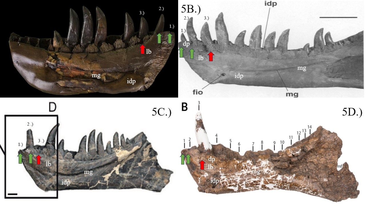
FMNH PR 2081 comes from Brochu (2003) (p. 41 Figure 40).Figure 5: T. rex dentaries in medial views. 5A: BHI 6439. 5B: FMNH PR 2081 from Brochu
(2003) (p. 42 Figure 41). 5C: BMNH R7994 from Dalman and Lucas (2017) (p. 24 Figure 10).
5D: RSM P2523.8 from Persons IV et al., (2019) (p. 668 Figure 18B). Numbers indicate the
position of the first three teeth in the dentary. The teeth in the dentary of RSM P2523.8 were
numbered by the authors of Persons IV et al., (2019). Green arrows indicate the lingual bar
covering the first two teeth/alveoli. The red arrows show that the lingual bar drops at the third
alveolus/tooth. The position of the lingual bar doesn’t change during ontogeny. Abbreviations:
“lb” is the lingual bar, “mg” is the Meckelian groove, “idp” is the interdentary plate, and “dp”
are the dental plates.Figure 6: Comparison between cf. Dryptosaurus specimen BMRP 2002.4.1 and T. rex specimen
BHI 6439 in dorsal view. 6A is the original photo. 6B is a close up, showing the 17th alveoli for
BMRP 2002.4.1 and the 13th alveoli for BHI 6439. Despite having a close tooth row length, BHI
6439 has fewer teeth than BMRP 2002.4.1. This demonstrates that young T. rex individuals
would’ve had a tooth count similar to the adults. The teeth in the T. rex specimen are also more
robust, while the teeth in the cf. Dryptosaurus specimen shows an extreme ziphodont
morphology. Photo belongs to Hiroshi Kato.Figure 7: Dentary of cf. Dryptosaurus specimen BMRP 2002.4.1 in dorsal view. 6A is the
original photo. 6B shows the number of teeth. 6C is the tooth count of BHI 6439 (also in dorsal
view). Tooth row length for BMRP 2002.4.1 is 31.5 cm. The tooth row length for BHI 6439 is 33
cm. Photo belongs to Hiroshi Kato.Figure 8: cf. Dryptosaurus specimen BMRP 2002.4.1 dentary from Dalman and Lucas (2017) (p.
24 Figure 9B and B’). 8A is the whole dentary. 8B is a close-up of the anterior tip. Numbers indicate the position of the teeth. Green arrows show that the lingual bar covers the first alveolus
only. Red arrows show that the lingual bar drops at the second alveolus, and continues past the
third alveolus. Abbreviations: “lb” is the lingual bar, “mg” is the Meckelian groove, and “idp” is
the interdentary plate.Figure 9: Close-ups of cf. Dryptosaurus specimen BMRP 2002.4.1 and T. rex specimen BHI
6439 (reversed). In 9A, the green arrow indicates that the lingual bar covers the first alveolus
only. The red arrows show that the lingual bar drops at the second alveolus, and past the third
one. In 9B, the green arrows show that the lingual bar covers the first alveoli and second tooth.
The red alveoli shows that the lingual bar drops at the third tooth. Abbreviations: “lb” is the
lingual bar, “mg” is the Meckelian groove, and “idp” is the interdentary plate.Figure 10: Close-ups of the anterior tips of basal tyrannosauroidea and tyrannosauridae dentaries
in medial views. Figures belong to Dalman and Lucas (2017). 10A is cf. Dryptosaurus specimen
BMRP 2002.4.1 (p. 24 Figure 9B’). 10B is the Alioramus altai holotype IGM 100/1844 (p. 23
Figure 6B) (reversed). 10C is Gorgosaurus specimen FPDM-V8062 (p. 19 Figure 2B). 10D is
Albertosaurus specimen TMP 1999.50.40 (p. 21 Figure 3B) (reversed). 10E is Bistahieversor
specimen NMMNH P-27469 (p. 29 Figure 9A’). The numbers indicate the placement of the
teeth/alveoli. The green arrows indicate that the first alveolus is covered by the lingual bar only.
The red arrows indicate that the second and third alveoli are not covered by the lingual bar.Figure 11: Skull and mandible bones of Appalachiosaurus holotype RMM 6670 compared to cf.
Dryptosaurus specimen BMRP 2002.4.1. Photo of Appalachiosaurus belongs to Joe Songer. 11A
shows the photo as is. 11B shows a close-up of the anterior tip of the dentary of
Appalachiosaurus in medial view (rotated). 11C shows the anterior portion of the dentary of
BMRP 2002.4.1 in medial view. Numbers indicate the position of the teeth/alveoli. Green arrows
indicate that the lingual bar covers the first alveolus only. The red arrows indicate that the lingual
bar drops past the second, third, and fourth alveoli. All basal tyrannosauroids and tyrannosaurids
share this characteristic.Figure 12: Close-ups of the anterior tip of the dentaries derived tyrannosaurinae taxa in medial
views. Figures belong to Dalman and Lucas (2017), and Fiorillo and Tykoski (2014). 12A is
Daspletosaurus torosus specimen TMP 2001.36.1 (Dalman and Lucas, 2017, p. 24 Figure 10A’).
12B is T. bataar specimen ZPAL MgD-I/5 (p. 24 Figure 10C’). 12C is Nanuqsaurus holotype
DMNH 21461 from Fiorillo and Tykoski (2014) (Figure 3) (reversed). 12D is Lythronax (or T.
argestes?) holotype UMNH VP 20200 (Dalman and Lucas, 2017, p. 24 Figure 10B’). 12E is
Zhuchengtyrannus (or T. magnus?) holotype ZCDM V0031 (p. 24 Figure 10E’). 12F is T. rex
specimen BMNH R7994 (p. 24 Figure 10D’). Numbers represent the tooth/alveoli placement.
Nanuqsaurus’ alveoli were numbered by the authors of Fiorillo and Tykoski (2014). Green
arrows indicate that the lingual bar covers the first two alveoli/teeth. Red arrows indicate that the
lingual bar drops at the third tooth/alveolus. Unlike the basal tyrannosauroids and tyrannosaurids
in Figure 11, all derived tyrannosaurinae here have the lingual bar covering the first two alveoli.Figure 13: Photos of an adult and baby T. rex specimen dentaries. 13A is a figure of the dentary
of the adult T. rex specimen NMMNH P-3698 from Larson (2008) (p. 41 Figure 1.24) (called
UMNH 110000 in the paper). 13B is a close-up of the anterior tip of the dentary in medial view.
13C is BHI 6439. Numbers indicate the position of the alveoli/teeth. Green arrows demonstrate
that the lingual bar covers the first two alveoli. The red arrows show that the lingual bar drops at
the third tooth/alveolus, and continues past the fourth tooth/alveolus. This characteristic is seen
in other T. rex specimens (see Figure 5), and other derived tyrannnosaurinae taxa (Figure 10).Figure 14: Daspletosaurus dentaries in medial views. 14A is an X-ray reconstruction of the
dentary of the cf. D. horneri hatchling MOR 268 in medial view from Funston et al., (2021)
(Figure 7). 14B is a close-up of the anterior portion of the dentary in medial view. 14C is the
anterior portion of the dentary of the D. torosus specimen TMP 2001.36.1. Numbers indicate the
position of the teeth/alveoli. The green arrows indicate that the lingual bar covers the first two
alveoli/teeth. The red arrows indicate that the lingual bar drops past the third alveolus. The
position of the lingual bar stays the same in different species of the same genus, regardless of
age.Figure 15: Sketch of the two different dentary morphologies seen within tyrannosauroidea in
medial views. 15A is the basal tyrannosauroidea and tyrannosauridae dentary morphology, with
the lingual bar covering the first alveolus only. This sketch was based on BMRP 2002.4.1. 15B is
the derived tyrannosaurinae dentary morphology, with the lingual bar covering the first two
alveoli. This sketch was based on BHI 6439. Numbers represent the amount of alveoli the lingual
bar covers. Abbreviations: “lb” is the lingual bar, “mg” is the Meckelian groove, and “idp” is the
interdentary plate. Sketch belongs to this author.
Tables:Table 1: Position of the lingual bar within multiple genera of tyrannosauroidea. Basal
tyrannosauroidea and tyrannosauridae taxa have the lingual bar covering the first alveoli only.
Derived tyrannosaurinae genera have the lingual bar covering the first two alveoli.Table 2: A comparison between the dentaries of T. rex specimen BHI 6439, and cf. Dryptosaurus
aquilunguis specimen BMRP 2002.4.1. BHI 6439 has 13 teeth that are robust in morphology, and a lingual bar covering the first two alveoli/teeth on the medial side of the dentary. BMRP
2002.4.1 has 17 teeth that have an extreme ziphodont morphology, and a lingual bar that covers
the first alveoli on the medial side of the bone. Both specimens have a close body size (6.7 to 7.0
meters), yet the two dentaries show major differences in morphology. The morphology of the
dentary for BMRP 2002.4.1 is closer to basal tyrannosauroidea and tyrannosauridae genera,
while BHI 6439 matches the morphology of derived tyrannosaurinae taxa.