Sunday, February 12, 2023

A Baby Tyrannosaurus rex Premaxillary, or First Maxillary, Tooth (2023)

Written on 1/17/23.

Link:
https://www.academia.edu/96575022/A_Baby_Tyrannosaurus_rex_Premaxillary_or_First_Maxillary_Tooth

                            A Baby Tyrannosaurus rex Premaxillary, or First Maxillary, Tooth

Abstract
In Dr. Kenneth Carpenter’s 1982 paper describing baby dinosaur dentaries and teeth, one 
tooth cataloged as UCMP 119853 seemed to have a morphology reminiscent of adult Tyrannosaurus rex specimens. After an extensive critique of the tooth, along with comments from other professional paleontologists (Professor Holtz, Jr., Sebastian Dalman, and Dr. Joshua B. Smith), this author believes that UCMP 119853 is a baby T. rex tooth that was situated in either the premaxillary (first or second), or the first maxillary, position. This author tends to lean more towards the premaxillary, but is still open to the possibility that the specimen is a first maxillary tooth. UCMP 119853 has a single carina that overlaps the tip of the tooth and is located in the labial and lingual positions of the crown, and the carina is denticulate. The morphology of UCMP 119853 differs from that of other young tyrannosauroid specimens that are categorized as either “Nanotyrannus,” or “juvenile T. rex specimens” (which this author categorizes as cf. Dryptosaurus aquilinguis). A comparison between UCMP 119853 and the premaxillary, and first maxillary, teeth from other tyrannosauroid genera showed that T. rex’s tooth morphology stayed consistent throughout the animal’s lifetime, and was close to that of the genus’ sister taxon Tyrannosaurus/Tarbosaurus bataar.

Figures:
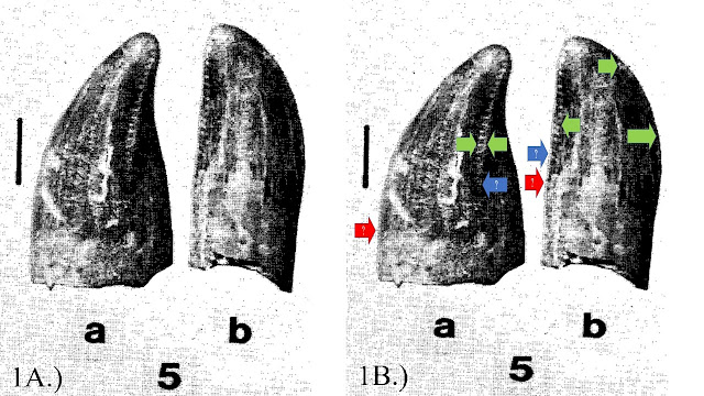
Figure 1: UCMP 119853. 1A.) The fossil as shown in Carpenter (1982). 5a is the lateral, and 5b is the posterior/lingual/distal view. 1B.) Arrows indicating the location, and endpoints, of the carina. Green arrows indicate the carina is located on the lateral (labial and lingual) sides of the tooth. The blue arrows indicate to the author where the carina ended. The red arrows indicate where the carine ended according to Dr. Smith:

Figure 2: A comparison of the posterior/lingual/distal views of 2A.) UCMP 119853, 2B) BHI 3033’s first premaxillary tooth, and 2C.) BHI 3033’s second premaxillary tooth. The carina are on the lateral (labial and lingual) sides of all the teeth. Interestingly, the curvature of UCMP 119853 is close to BHI 3033’s second premaxillary tooth in particular. Photos of BHI 3033’s teeth were provided by Dr. Smith:
Figure 3: 
Comparisons in lateral view of 3A.) UCMP 119853, 3B.) first premaxillary tooth of BHI 3033, and 3C.) first maxillary tooth of BHI 3033. Green arrows indicate the location of the carina. Lines indicate the exterior mesial outline of the teeth. You can see that the first maxillary tooth is more elongated and thin (red lines), compared to the curvier premaxillary teeth (green lines). Photos of BHI 3033’s teeth were provided by Dr. Smith:
Figure 4: 
Posterior/lingual/distal views of 4A.) UCMP 119853, and 4B.) BHI 3033’s first maxillary tooth (flipped). Green arrows indicate the location of the carina. The overall curvature of the BHI 3033’s first maxillary tooth seems to be more linear compared to UCMP 119853’s, especially on the right side of each tooth. Photo of BHI 3033’s tooth was provided by Dr. Smith:
Figure 5: 
Lateral views of 5A.) UCMP 119853, and 5B.) first premaxillary tooth of MOR 008. Carina are indicated by green arrows. The overall shape of both teeth seem to match. Photo of MOR 008’s tooth was provided by Dr. Smith:
Figure 6: 
Lateral views of 6A.) UCMP 119853 and 6B.) TD-13-251 (rotated and flipped) from Stein (2021). Green arrows indicate carina in lateral (labial and lingual) view. Blue arrows show that the carina could have stopped midpoint on the crown of UCMP 119853 and TD-13-251. Photos of BHI 3033’s teeth were provided by Dr. Smith:
Figure 7: 
Figure 1 from Hendrickx et al., (2019) showing the views of the teeth and carina locations. Vocabulary words used here were used in this paper:
Figure 8: 
UCMP 124406. 8A.) The specimen as shown in Carpenter (1982). 8B.) The specimen with arrows. Red arrows show the carinae located on the distal end of the tooth. Purple arrows indicate the posterior/distal ventral ridge:
Figure 9: Comparisons between 9A.) UCMP 124406, 9B.) LACM 28471 from Molnar (1978), 9C.) FMNH PR 2902 from Gates et al., (2015), and 9D.) YPM 296 from Marsh (1892). Red arrows indicate the carinae on the posterior/distal end. Purple arrows indicate the posterior/distal vertical ridge. The morphology of the teeth stays consistent during the animal’s growth:
Figure 10: Comparisons between 9A.) UCMP 124406, 9B.) LACM 28471 from Molnar (1978), 9C.) FMNH PR 2902 from Gates et al., (2015), and 9D.) YPM 296 from Marsh (1892). Red arrows indicate the carinae on the posterior/distal end. Purple arrows indicate the posterior/distal vertical ridge. The morphology of the teeth stays consistent during the animal’s growth:
Figure 11: A comparison between 11A.) UCMP 119853, and (11B.) UCMP 124406. UCMP 119853’s carina is on the lateral (labial and lingual) sides (green arrows) that are serrated, and lacks a vertical ridge on the posterior/distal end of the crown. UCMP 124406 has distal carinae (red arrows) that lack serrations, and a distal vertical ridge (purple arrows) on the distal end of the crown:
Figure 12: 12A.) Figure 17 from Stein (2021). 12B.) Close-up of TD-13-251 and TD-13-247, comparing their sizes to each other. The two specimens are close in size, yet have different morphologies:
Figure 13: Comparison between 13A.) UCMP 119853, 13B.) TD-13-251, and 13C.) BHI 3033’s first premaxillary tooth. The green arrows represent the carina on the lateral (labial and lingual) sides. Red and blue arrows indicate possible endings of the carina. The morphology of the teeth stays consistent during the animal’s growth:
Figure 14: 
Comparison between 14A.) UCMP 119853, and 14B.) 2-3-year old T. bataar specimen MPC-D 107/7’s premaxillary teeth in labiodistal view (Tsuihiji et al., 2011, Figure 6C-D). The green arrows indicate the serrated carina in lateral (labial and lingual) view. In 14B.), the blue semi-circle and bar show the distal side of MPC-D 107/7’s premaxillary teeth:
Figure 15: 
The two teeth morphotypes from the late Maastrichtian of North America. A, C, and E is the cf. Dryptosaurus aquilinguis premaxillary tooth. B, D, and E is the T. rex premaxillary tooth. Vocabulary comes from Hendrickx et al., (2019). Illustration belongs to this author:
Tables:
Table 1: 
T. rex and cf. Dryptosaurus aquilinguis premaxillary tooth lengths and morphologies. The results indicated that the morphologies of the teeth in the two genera stayed consistent, aside from an increase in size during maturity. “(C)” means crown height measurement, and “(T)” means total tooth height measurement. Note: This author obtained a length of 3 cm for YPM 296, but Ford and Chure (2001) gave 2.9 cm (Table 1). This was discovered after the author already measured the specimen:
Table 2: Tyrannosauroid premaxillary tooth morphologies. The results showed that the morphology of the cf. Dryptosaurus aquilinguis premaxillary teeth were closer to the other basal tyrannosauroids than to T. rex’s. T. rex’s premaxillary tooth morphology was closer to T. bataar’s than the other taxa listed:
Friday, February 10, 2023

"Nanotyrannus" is Dryptosaurus: An Abstract (2022)

Written from 8/3/22-2/9/23.

Link:
https://www.academia.edu/96574784/Nanotyrannus_is_Dryptosaurus_An_Abstract

                                                “Nanotyrannus” is Dryptosaurus: An Abstract

This is an updated version of an abstract written in 2022.

Dryptosaurus aquilunguis is a tyrannosauroid from the late Maastrichtian of Eastern North America, also known as Appalachia. So far, only one good specimen, the holotype ANSP 9995, has been found for the genus. A few teeth have been assigned, but no relatively complete specimens have been described yet. However, after an exhaustive examination of the controversial “Nanotyrannus”/juvenile Tyrannosaurus rex specimens, this author is going to introduce a new hypothesis: Dryptosaurus lived in Appalachia and Laramidia towards the end of the Maastrichtian. The tyrannosauroid specimens previously labeled as “Nanotyrannus” are either cf. Dryptosaurus aquilinguis, or a sister taxon to Dryptosaurus and the two genera form a clade within tyrannosauroidea. This author tends to lean more towards “Nanotyrannus” being cf. Dryptosaurus aquilinguis. Both Dryptosaurus and “Nanotyrannus” lived during the same time. Numerous publications have suggested that Laramidia and Appalachia reconnected when the Western Interior Sea subsided in the Maastrichtian. Both Laramidia and Appalachia seemed to have had similar fauna: lambeosaurs, ceratopsians, and mosasaurs. Ceratopsids, in particular, were thought to have not existed in Appalachia. However, a ceratopsian tooth has been found in the Maastrichtian-aged Owl Creek Formation, which is in Appalachia. If animals in Laramidia can be found in Appalachia, and vice versa, then Dryptosaurus could’ve migrated into Laramidia. Dryptosaurus and “Nanotyrannus” share many physical characteristics, and the “Nanotyrannus” specimens have many traits not seen in T. rex. A few traits include:

1. Nanotyrannus” had a first maxillary tooth that was incisiform. This morphology is also seen in premaxillary teeth. The “Nanotyrannus” specimens lack serrations, and have two carinae on the distal side of the premaxillary, and first maxillary, teeth. This trait is not present in T. rex. Specimen UCMP 119853, a 8-mm long tooth that likely belongs in the premaxillary position, has one serrated carina that is located on the labial and lingual positions of the crown. This morphology is seen in larger T. rex specimens, showing that T. rex’s tooth morphology didn’t change during ontogeny. More than likely, the first maxillary tooth in young T. rex specimens wasn’t similar to the “Nanotyrannus” or Dryptosaurus specimens.

2. The maxillary, and perhaps dentary, teeth in both Dryptosaurus and “Nanotyrannus” were pinched on the labial and lingual sides in cross-section, and the denticle morphology was “hook-shaped.” T. rex’s cross-sections were oval-shaped, and the denticles were square/rectangular in shape. 

3. The dentary in the “Nanotyrannus” specimens had the lingual bar covering the first alveoli on the medial side, as seen in more basal tyrannosauroids like Appalachiosaurus, Gorgosaurus, Albertosaurus, and Bistahieversor. Since Dryptosaurus is a basal tyrannosauroid as well, it’s more than likely that it had this trait too. The derived tyrannosaurinae taxa T. rex, T. bataar, and Daspletosaurus had the lingual bar covering the first two alveoli. It seems that the lingual bar’s position can help differentiate between basal tyrannosauroids and tyrannosaurids from the derived tyrannosaurinae. The 3-4-year old T. rex specimen “Baby Bob” had the lingual bar covering the first two alveoli, as seen in the adult specimens. This alone places the “Nanotyrannus” specimens in basal tyrannosauroids or tyrannosaurids, not tyrannosaurinae.

4. The tooth count in the dentaries of baby-juvenile specimens of T. rex were the same as the adults, and the maximum count seems to be fifteen. This is also seen in T. bataar. The 3-4-year old specimen “Baby Bob” had 12 teeth in its dentary, as in the slightly larger baby/juvenile specimen BHI 6439. The 18-year old specimen BHI 3033 had 13. The 23-year old specimen CM 79057 ("Samson"), and PARC-TD-11-094/FDM-xx? (perhaps a subadult/adult individual), had 15. “Nanotyrannus” had 16-17. There is no sign of tooth loss during ontogeny.

5. The “Nanotyrannus” specimens have a non-incisiform, conical-shaped first dentary tooth with carina/carinae on the mesial and distal positions. This is also seen in Albertosaurus, but not in Gorgosaurus. T. rex’s first dentary tooth has the same morphology seen in the premaxillary teeth (incisiform, with a serrated carina on the labial and lingual faces). This is seen in the 3-4-year old “Baby Bob,” and the the 18-year old BHI 3033. The first dentary tooth never changed morphology as the T. rex individual aged, just like the premaxillary teeth.

6. Both genera have similar caudal vertebrae morphologies. In terms of the vertebrae count, it seems to be about 25 or so, as suggested by Cope. T. rex and Tarbosaurus/Tyrannosaurus bataar have 40 or more caudal vertebrae, and this is also seen in the young T. bataar specimen PIN 552-2. Whether or not this number is permanent doesn’t concern this author, but the morphology of the vertebrae does. From the middle to the distal portion of the tail, both Dryptosaurus and “Nanotyrannus” have elongated caudals that also lack transverse processes. T. rex’s caudals became shorter up to the very last caudal, and most of the caudals up until the last distal members had transverse processes. The middle-distal caudals were longer in Dryptosaurus than T. rex’s, and “Nanotyrannus’” looks to be the same.

7. Morphology of the arms of both genera are identical. The deltopectoral crest is positioned lower from the proximal end of the humerus, and faces different directions when viewed in multiple positions than in T. rex’s and other tyrannosauroids. For example, in proximal view, the deltopetoral crest in Dryptosaurus and “Nanotyrannus” faced anteriorly compared to the laterally-facing crest in T. rex. The manual phalanx 1-1 of Dryptosaurus and “Nanotyrannus” are extremely elongated, and this is an autapomorphy of Dryptosaurus. However, Megaraptor has this as well. Other tyrannosauroids like T. rex (9.85 cm), Gorgosaurus (9.8 cm), Albertosaurus (8.5 cm), and T. bataar (5.4 cm) have smaller manual phalanx 1-1 bones compared to Dryptosaurus’ (16 cm) or “Nanotyrannus’.” The manual unguals of the two genera are large and comparable in morphology and size, contra to T. rex’s and T. bataar’s short manual unguals.

8. Both Dryptosaurus and “Nanotyrannus” were agile tyrannosauroids throughout their biogenic existence, based on their hindlimb proportions. The tibia in the “Nanotyrannus” specimens are longer than the femur. The Dryptosaurus holotype seems to have a femur and tibia that were either equal in length, or the tibia was slightly longer than the femur. Dryptosaurus’ tibia is slightly eroded, so it might’ve been longer in life. Regardless, the femur and tibia of the “Nanotyrannus” specimens may have grown to become equal in length when they reached maturity, as possibly seen in Dryptosaurus. Or, both Dryptosaurus and “Nanotyrannus” had a tibia longer than the femur. Baby-juvenile specimens of T. rex and T. bataar had femora and tibiae that were about equal in length to each other, while the adults had longer femora. The 3-4-year old ”Baby Bob” demonstrates this. Body mass could account for this. Dryptosaurus and “Nanotyrannus” were small-medium-sized tyrannosauroids, so having a longer tibia, or a femur and tibia of equal length, could’ve meant a speedier lifestyle for the two genera. Another example of this is the adult Qiazhousaurus/Alioramus sinensis specimen, which also had a longer tibia compared to its femur. Larger adult tyrannosauroids had more massive bodies, which resulted in larger femora to support their weight. Indeed, baby-juvenile specimens of large tyrannosauroids had the femora and tibiae at about an equal length, but this changed during ontology. This doesn’t seem to be the case for Dryptosaurus, nor “Nanotyrannus.”

The fact that there are baby-juvenile specimens of T. rex that have traits not seen in the “Nanotyrannus” specimens, but are seen in the older specimens, show that “Nanotyrannus” doesn’t belong in the genus Tyrannosaurus. The stratigraphic and geographic correlations, as well as the similarities in characteristics with Dryptosaurus, show that “Nanotyrannus” is a more basal tyrannosauroid than a derived tyrannosaurinae. Further analyses will be conducted in the future to explore these characteristics in more explicit detail. 

Thursday, February 9, 2023

My Researchgate and Academia Accounts Update:

I've decided to delete my Researchgate account. I had a misunderstanding with the "Institution" section. I am just an undergraduate student at UMD, and I'm not associated with any program at the university. I did, however, create an Academia account to upload my work to. All of my future papers will be posted there. I already uploaded my abstract and first paper to the account.

My papers:
1. "Nanotyrannus" is Dryptosaurus: An Abstract (2022):
https://www.academia.edu/96574784/Nanotyrannus_is_Dryptosaurus_An_Abstract
2. A Baby Tyrannosaurus rex Premaxillary, or First Maxillary, Tooth (2023):
https://www.academia.edu/96575022/A_Baby_Tyrannosaurus_rex_Premaxillary_or_First_Maxillary_Tooth