Written on 1/17/23.
Link:
https://www.academia.edu/96575022/A_Baby_Tyrannosaurus_rex_Premaxillary_or_First_Maxillary_Tooth
A Baby Tyrannosaurus rex Premaxillary,
or First Maxillary, Tooth
Abstract
In Dr. Kenneth Carpenter’s 1982 paper describing baby dinosaur dentaries and teeth, one tooth cataloged as UCMP 119853 seemed to have a morphology reminiscent of adult
Tyrannosaurus rex specimens. After an extensive critique of the tooth, along with comments
from other professional paleontologists (Professor Holtz, Jr., Sebastian Dalman, and Dr. Joshua
B. Smith), this author believes that UCMP 119853 is a baby T. rex tooth that was situated in
either the premaxillary (first or second), or the first maxillary, position. This author tends to lean
more towards the premaxillary, but is still open to the possibility that the specimen is a first
maxillary tooth. UCMP 119853 has a single carina that overlaps the tip of the tooth and is
located in the labial and lingual positions of the crown, and the carina is denticulate. The
morphology of UCMP 119853 differs from that of other young tyrannosauroid specimens that
are categorized as either “Nanotyrannus,” or “juvenile T. rex specimens” (which this author
categorizes as cf. Dryptosaurus aquilinguis). A comparison between UCMP 119853 and the
premaxillary, and first maxillary, teeth from other tyrannosauroid genera showed that T. rex’s
tooth morphology stayed consistent throughout the animal’s lifetime, and was close to that of the
genus’ sister taxon Tyrannosaurus/Tarbosaurus bataar.
Figures:
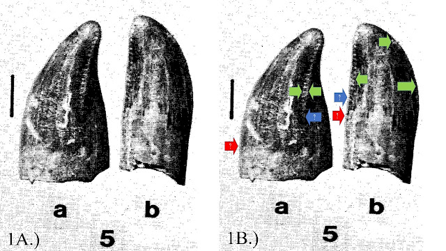
Figure 1: UCMP 119853. 1A.) The fossil as shown in Carpenter (1982). 5a is the lateral, and 5b
is the posterior/lingual/distal view. 1B.) Arrows indicating the location, and endpoints, of the
carina. Green arrows indicate the carina is located on the lateral (labial and lingual) sides of the
tooth. The blue arrows indicate to the author where the carina ended. The red arrows indicate
where the carine ended according to Dr. Smith:
Figure 2:
A comparison of the posterior/lingual/distal views of 2A.) UCMP 119853, 2B) BHI
3033’s first premaxillary tooth, and 2C.) BHI 3033’s second premaxillary tooth. The carina are
on the lateral (labial and lingual) sides of all the teeth. Interestingly, the curvature of UCMP
119853 is close to BHI 3033’s second premaxillary tooth in particular. Photos of BHI 3033’s
teeth were provided by Dr. Smith:Figure 3: Comparisons in lateral view of 3A.) UCMP 119853, 3B.) first premaxillary tooth of
BHI 3033, and 3C.) first maxillary tooth of BHI 3033. Green arrows indicate the location of the
carina. Lines indicate the exterior mesial outline of the teeth. You can see that the first maxillary
tooth is more elongated and thin (red lines), compared to the curvier premaxillary teeth (green
lines). Photos of BHI 3033’s teeth were provided by Dr. Smith:Figure 4: Posterior/lingual/distal views of 4A.) UCMP 119853, and 4B.) BHI 3033’s first
maxillary tooth (flipped). Green arrows indicate the location of the carina. The overall curvature
of the BHI 3033’s first maxillary tooth seems to be more linear compared to UCMP 119853’s,
especially on the right side of each tooth. Photo of BHI 3033’s tooth was provided by Dr. Smith:Figure 5: Lateral views of 5A.) UCMP 119853, and 5B.) first premaxillary tooth of MOR 008.
Carina are indicated by green arrows. The overall shape of both teeth seem to match. Photo of
MOR 008’s tooth was provided by Dr. Smith:Figure 6: Lateral views of 6A.) UCMP 119853 and 6B.) TD-13-251 (rotated and flipped) from
Stein (2021). Green arrows indicate carina in lateral (labial and lingual) view. Blue arrows show
that the carina could have stopped midpoint on the crown of UCMP 119853 and TD-13-251.
Photos of BHI 3033’s teeth were provided by Dr. Smith:Figure 7: Figure 1 from Hendrickx et al., (2019) showing the views of the teeth and carina
locations. Vocabulary words used here were used in this paper:Figure 8: UCMP 124406. 8A.) The specimen as shown in Carpenter (1982). 8B.) The specimen
with arrows. Red arrows show the carinae located on the distal end of the tooth. Purple arrows
indicate the posterior/distal ventral ridge:Figure 9: Comparisons between 9A.) UCMP 124406, 9B.) LACM 28471 from Molnar (1978),
9C.) FMNH PR 2902 from Gates et al., (2015), and 9D.) YPM 296 from Marsh (1892). Red
arrows indicate the carinae on the posterior/distal end. Purple arrows indicate the posterior/distal
vertical ridge. The morphology of the teeth stays consistent during the animal’s growth:Figure 10: Comparisons between 9A.) UCMP 124406, 9B.) LACM 28471 from Molnar (1978),
9C.) FMNH PR 2902 from Gates et al., (2015), and 9D.) YPM 296 from Marsh (1892). Red
arrows indicate the carinae on the posterior/distal end. Purple arrows indicate the posterior/distal
vertical ridge. The morphology of the teeth stays consistent during the animal’s growth:Figure 11: A comparison between 11A.) UCMP 119853, and (11B.) UCMP 124406. UCMP
119853’s carina is on the lateral (labial and lingual) sides (green arrows) that are serrated, and
lacks a vertical ridge on the posterior/distal end of the crown. UCMP 124406 has distal carinae
(red arrows) that lack serrations, and a distal vertical ridge (purple arrows) on the distal end of
the crown:Figure 12: 12A.) Figure 17 from Stein (2021). 12B.) Close-up of TD-13-251 and TD-13-247,
comparing their sizes to each other. The two specimens are close in size, yet have different
morphologies:Figure 13: Comparison between 13A.) UCMP 119853, 13B.) TD-13-251, and 13C.) BHI 3033’s
first premaxillary tooth. The green arrows represent the carina on the lateral (labial and lingual)
sides. Red and blue arrows indicate possible endings of the carina. The morphology of the teeth
stays consistent during the animal’s growth:Figure 14: Comparison between 14A.) UCMP 119853, and 14B.) 2-3-year old T. bataar
specimen MPC-D 107/7’s premaxillary teeth in labiodistal view (Tsuihiji et al., 2011, Figure
6C-D). The green arrows indicate the serrated carina in lateral (labial and lingual) view. In 14B.),
the blue semi-circle and bar show the distal side of MPC-D 107/7’s premaxillary teeth:Figure 15: The two teeth morphotypes from the late Maastrichtian of North America. A, C, and
E is the cf. Dryptosaurus aquilinguis premaxillary tooth. B, D, and E is the T. rex premaxillary
tooth. Vocabulary comes from Hendrickx et al., (2019). Illustration belongs to this author:Tables:
Table 1: T. rex and cf. Dryptosaurus aquilinguis premaxillary tooth lengths and morphologies.
The results indicated that the morphologies of the teeth in the two genera stayed consistent, aside
from an increase in size during maturity. “(C)” means crown height measurement, and “(T)”
means total tooth height measurement. Note: This author obtained a length of 3 cm for YPM
296, but Ford and Chure (2001) gave 2.9 cm (Table 1). This was discovered after the author
already measured the specimen:Table 2: Tyrannosauroid premaxillary tooth morphologies. The results showed that the
morphology of the cf. Dryptosaurus aquilinguis premaxillary teeth were closer to the other basal
tyrannosauroids than to T. rex’s. T. rex’s premaxillary tooth morphology was closer to T. bataar’s
than the other taxa listed:


.png)


.jpg)

